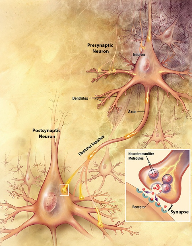
FIGURE 1.
Anatomy of two representative neurons in the brain and a synapse between them. Path of electrical current indicated with yellow arrows. Inset, close-up view of the synapse. Illustration adapted from Alzheimer’s Disease Education and Referral Center, National Institutes on Aging, U.S. National Institutes of Health (www.nia.nih.gov/Alzheimers/Publications/UnravelingtheMystery).

