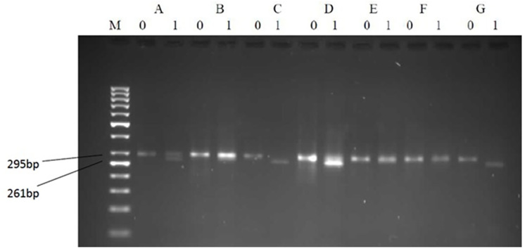
Figure 2.
Representative patterns on agarose gel electrophoresis illustrating the restriction enzyme-based detection of precore mutants and variants. Each analysis is represented by two lanes: (1) the left without and (2) the right with a restriction enzyme Bsu36I added. B and E show TGG (wild-type); C, D, and G show TAG (mutant); and A shows a mixture of TGG/TAG at codon 28; M: Marker 50bp.

