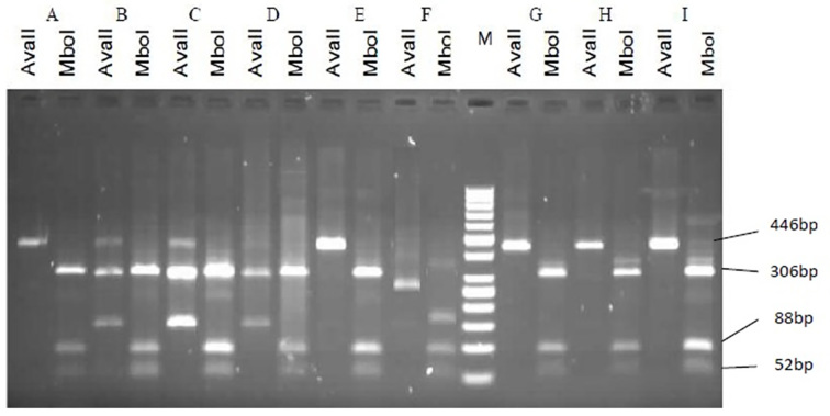
Figure 4.
Representative patterns on agarose gel electrophoresis illustrating the restriction enzyme-based detection of HBV genotypes. Each analysis is represented by two lanes: (1) the left with a restriction enzyme AvaII; (2) the right with a restriction enzyme MboI added. M: Marker 50bp. First pattern: Samples A, E, H, I, and G, which are matched with a D2 pattern of Lindh patterns that were cut into three fragments of 52nt, 88nt, and 306nt by MboI and had no excision site for AvaII (446bp). Second pattern: Sample F, which is matched with Ddel pattern of Lindh patterns that were cut into three fragments of 52nt, 88nt, and 123nt by MboI and had no excision site for AvaII, which produced a 263bp fragment. The third pattern belonged to samples B, C, and D, which were not matched with any of the Lindh patterns.

