
Figure 2.
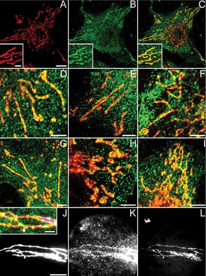
Confocal and wide-field microscopical data of ECV 304, HeLa, and SK-BR-3 cells labeled with MitoTracker (red) and anti-BRCA1-FITC/Alexa Fluor 488 antibodies (green). PFA-fixed ECV 304 cells stained with Ab-5 (B and C) and Ab-1 (E) and methanol-fixed ECV 304 cells stained with Ab-4 (D), Ab-C (F), and Ab-D (G) show granular BRCA1 cytoplasmic staining and colocalization of linear MitoTracker staining with linear BRCA1 staining results in yellow. (A) Single MitoTracker staining, overview. (B) Single Ab-5 staining, overview. (C) Merge of A and B. Insets in A–C represent a higher magnification. Double labeling of PFA-fixed HeLa (H) and SK-BR-3 (I) cells with MitoTracker and Ab-5 again shows yellow colocalization of both labels. Wide-field microscopical data shows colocalization of MitoTracker (J), Ab-5 (K), and mtDNA DAPI (L) on PFA-fixed ECV 304 cells, resulting in white foci (J, inset: higher magnification merge of J–L). Bars, 3 μm in insets; 5 μm in the other panels.