
Figure 3.
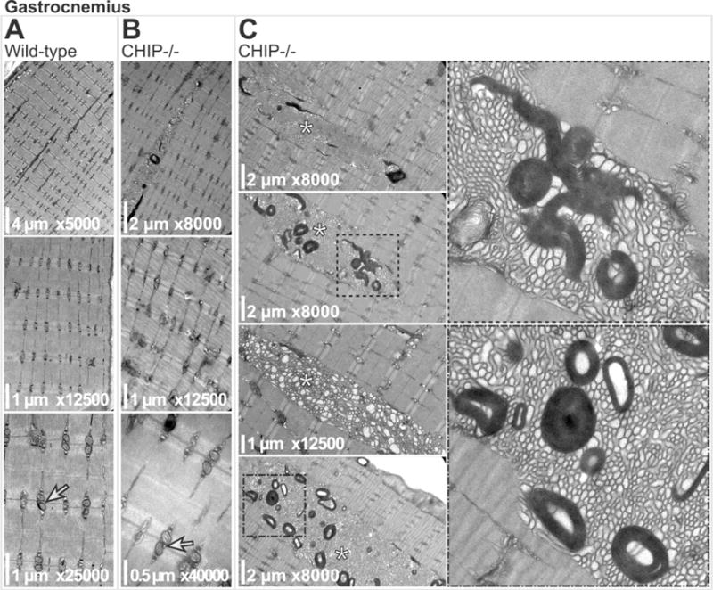
Transmission electron micrographs of gastrocnemius muscle sarcomeres from (A) wild-type and (B) CHIP−/− mice. Longitudinal doublet mitochondria are indicated (arrows in A, B, lower panels). (C) Micrographs of CHIP−/− gastrocnemius muscle sarcomeres with tubular aggregates (indicated by * in left panels). Boxed regions are magnified (right panels) highlighting the with laminar bodies