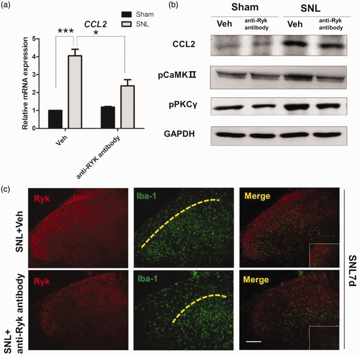
Figure 7.
Ryk activation after SNL surgery induced increased secretion of CCL2 through CaMKII/ PKCγ pathway. (a) Immunohistochemistry shows the expression of Ryk (red) and Iba-1(green) seven days after SNL surgery with and without the administration of anti-Ryk. (Scale bar, 100 µm). (b) Western blot analysis of CCL2, phospho-CaMKII, and phospho-PKCγ for proteins harvested from spinal cord seven days after surgery. Data are shown as mean ± SEM. *p < 0.05, ***p < 0.001 versus control in the corresponding group. (c) Immunohistochemical staining showed that in spinal cord with SNL surgery, Ryk (red) highly expressed area could attract microglia invasion (Iba-1, green), which is lamina II area of the dorsal horn. For microglia (Iba-1 positive) in spinal cord, blocking Ryk receptors with anti-Ryk results in the reduction of microglia invasion into lamina II area of the dorsal horn on post-operative day 7.

