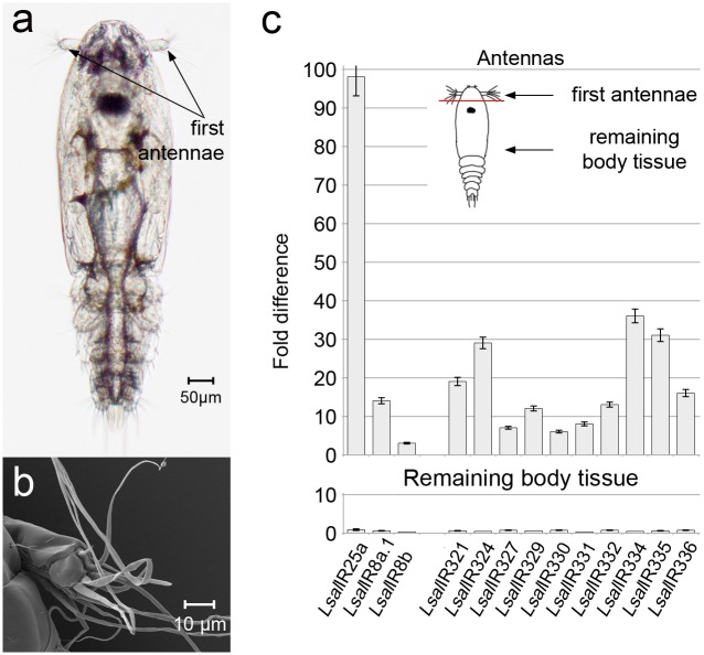
Fig 1. Copepodid, infectious stage of L.salmonis and antenna-specific expression of IRs: Co-receptors and antennal IRs.
(a). Free living, infectious stage of L. salmonis with marked sensory organs—first antennae. (b). Close up of copepodid’s first antenna in SEM. Long sensilla are located mainly on the most distal segment. (c). Expression of co-receptors and antennal IRs in antennae and remaining body tissue. Expression levels are given as a fold difference compared to intact animal. Bars indicate standard deviation. Analysis was performed on three parallels, where each antennal sample consisted of antennae pairs dissected from 500 animals, and the remaining body samples consisted of 100 dissected animals. On the right top, drawing of copepodid showing first antennae dissection for expression analysis.

