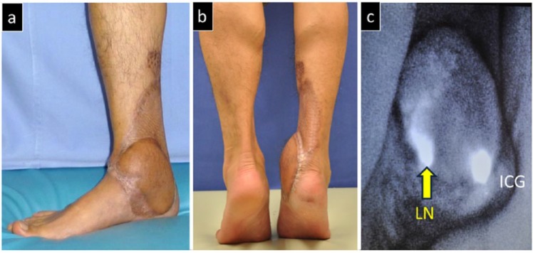
Figure 3.
Post-operative view of Case 1. (a) Complete epithelialization of the wound is observed after a 10-month post-operative follow-up. (b) The functional impairment in the ankle joint is kept to a minimum. The patient is able to stand on tiptoes. (c) Post-operative ICG imagining shows accumulation of ICG in the transplanted lymph nodes, thus confirming the attachment of the nodes.
ICG: indocyanine green; LN: lymph node.

