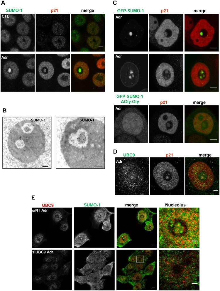
Fig 1. p21 and SUMO-1 colocalize in the disrupted nucleolus upon DNA damage.
A) Immunodetection of endogenous SUMO-1 (green) using anti-SUMO-1 mouse antibody and p21 (red) using anti-p21 rabbit antibody in control (CTL) or treated with Adr for 48 hours (Adr) HCT116 cells. Scale bar: 5μm. B) Immunogold electronic microscopy of SUMO-1 showing the presence of SUMO-1 in the INoB HCT116 cells treated with Adr for 24 hours. Scale Bar: 0.5μm. C) Immunostaining of endogenous p21 (red) in GFP-SUMO-1 (two representative cells are shown) or GFP-SUMO-1ΔGly-Gly transfected cells treated 24h with Adr. Scale bar: 5μm. D) Immunostaining of p21 (red) and UBC9 (green) (rabbit polyclonal antibody) in 24-h Adr-treated HCT116 cells. Scale bar: 5μm. E) Immunostaining of SUMO-1 (green) and UBC9 (red) (rabbit monoclonal antibody) of 24-h Adr-treated HCT116 cells transfected with non-targeting (siNT) or UBC9 (siUBC9). Scale bar: 5 μm.

