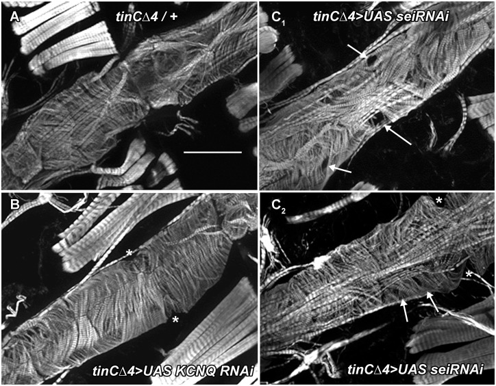
Fig 9. Cardiac-specific sei KD causes myofibrillar disarray and thinning.
(A) Phalloidin staining of F-actin in hearts from 3 week old flies reveals the circumferential myofibrillar structure in control (tinCΔ4Gal4/+). Scale bar is 50μm. (B) Hearts for 3 week old KCNQ KD flies (tinCΔ4Gal4>UASKCNQ RNAi) also show a normal circumferential myofibrillar pattern. (C) Cardiac-specific KD of sei results in myofibrillar disarray and gaps (arrows). * denotes the position of ostia.

