Figure 2.

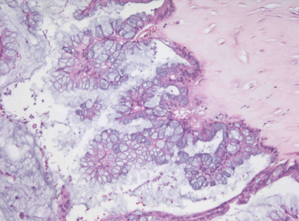
The section reveals papillary hyperplasia of mucinous lining epithelium with stratification and atypism (H&E stain, ×250).

The section reveals papillary hyperplasia of mucinous lining epithelium with stratification and atypism (H&E stain, ×250).