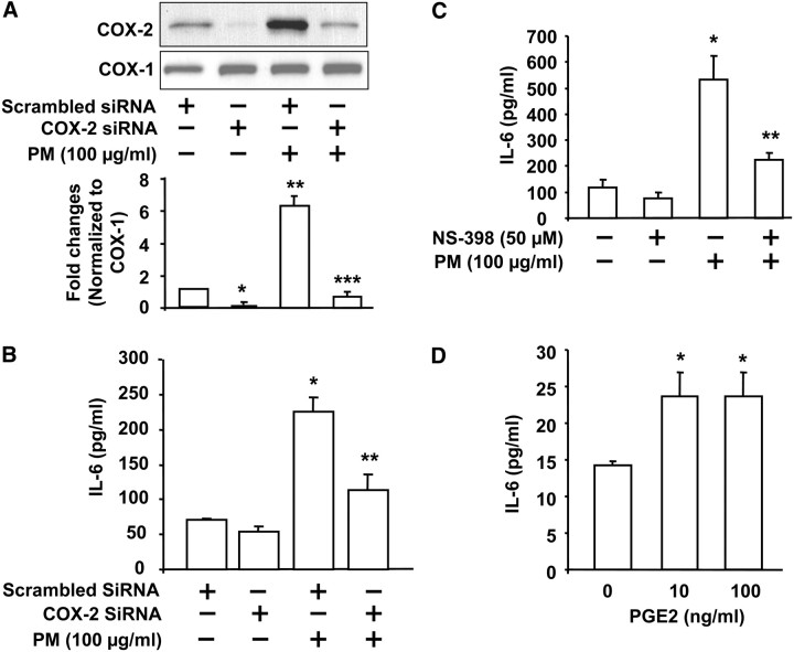
Figure 10.
Involvement of COX-2 and PGE2 in Baltimore PM–induced IL-6 secretion. In A and B, HBEpCs grown on 35-mm dishes to approximately 50% confluence were transfected with scrambled siRNA (50 nM) or COX-2 siRNA (50 nM) for 72 hours before challenge with vehicle or vehicle plus Baltimore PM (100 μg/ml) for 24 hours. In A, cell lysates (20 μg proteins) were subjected to SDS-PAGE, and Western blotted with anti–COX-2 or –COX-1 antibodies. Shown are representative blots and quantitative analyses from three independent experiments. * and ** significantly different from scrambled siRNA-transfected cells exposed to vehicle (P < 0.01); *** significantly different from COX-2 siRNA-transfected cells exposed to PM (P < 0.01). In B, media from A were collected and analyzed for IL-6 by ELISA. Values are mean ± SD from three independent experiments. *Significantly different from scrambled siRNA-transfected cells exposed to vehicle (P < 0.01); **significantly different from scrambled siRNA-transfected cells exposed to Baltimore PM (P < 0.01). In C, HBEpCs grown to approximately 90% confluence were pretreated with COX-2 inhibitor, NS-398 (50 μM) for 1 hour, cells were challenged with vehicle or vehicle plus Baltimore PM (100 μg/ml) for 24 hours, and media were analyzed for IL-6 by ELISA. Values are mean ± SD from three independent experiments. *Significantly different from cells challenged with vehicle (P < 0.05); **significantly different from cells exposed to Baltimore PM (P < 0.01). In D, PGE2 (10, and 100 ng/ml) was added to HBEpCs grown on 35-mm dishes for 6 hours, media were collected, and analyzed for IL-6 by ELISA. Values are mean ± SD from three independent experiments. *Significantly different from cells exposed to vehicle (P < 0.05).

