Figure 3.
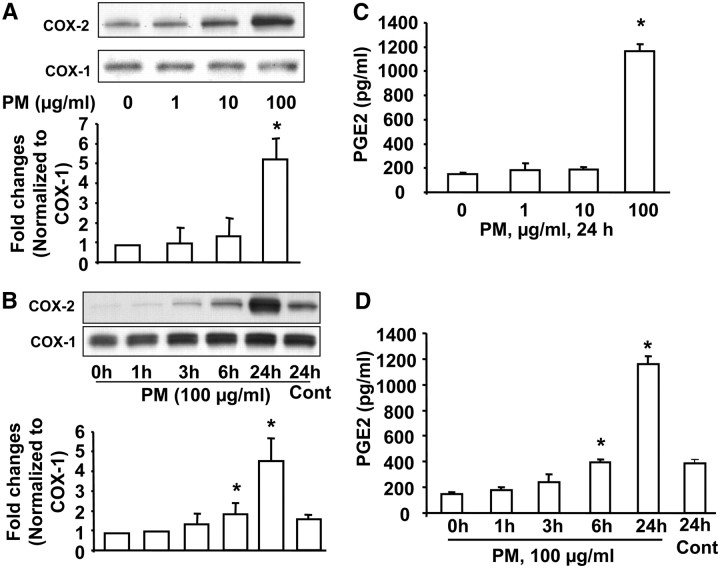
Baltimore PM induces cyclooxygenase (COX)-2 expression and prostaglandin (PG)E2 release. In A and C, HBEpCs grown to approximately 90% confluence were treated with varying concentrations of Baltimore PM (1, 10, and 100 μg/ml) for 24 hours. (A) Cell lysates (20 μg proteins) were subjected to SDS-PAGE and Western blotted with anti–COX-1 and –COX-2 antibodies. Shown are representative blots from three independent experiments. Quantitative analyses from three independent experiments (mean ± SD). *P < 0.05 versus vehicle. (C) Total PGE2 released in the medium was quantified by ELISA. Values are mean ± SD of three independent experiments in triplicate and expressed as pg of PGE2/ml of medium. *Significantly different from vehicle-treated cells (P < 0.01). In B and D, HBEpCs (∼ 90% confluence) were exposed to Baltimore PM (100 μg/ml) for 1, 3, 6, and 24 hours. At each time point, media were collected and cell lysates were prepared. (B) Cell lysates (20 μg proteins) were subjected to SDS-PAGE and Western blotted for COX-1 and COX-2. Shown are representative blots from three independent experiments. Quantitative analyses from three independent experiments (mean ± SD). *P < 0.05 versus 0 hours. (D) Media from the various time points were analyzed for PGE2 release by ELISA. Values are mean ± SD of three independent experiments in triplicate and expressed as pg of PGE2/ml of medium. *Significantly different from vehicle-treated cells (P < 0.01).

