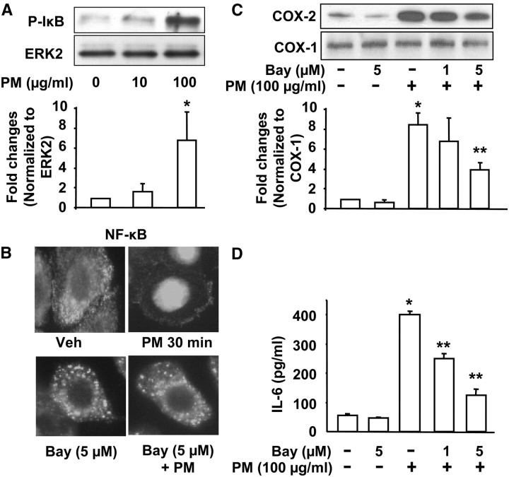
Figure 7.
Baltimore PM-induced COX-2 expression and IL-6 secretion via NF-κB. In A, HBEpCs grown in 35-mm dishes to approximately 95% confluence were starved in basal EBM medium without any growth factors for 3 hours, and then challenged with Baltimore PM (10 and 100 μg/ml) for 15 minutes. Cell lysates (20 μg proteins) were subjected to SDS-PAGE and Western blotted with phospho-IkB and ERK2 antibodies. Shown are representative blots and quantitative analyses from three independent experiments (mean ± SD). *P < 0.05 versus vehicle. **P < 0.05 versus PM challenge. In B, HBEpCs in glass coverslips (∼ 95% confluence) were pretreated with Bay compound (5 μM) for 60 minutes, cells were challenged with vehicle or vehicle plus Baltimore PM (100 μg/ml) for 15 minutes. Cells were washed, fixed, permeabilized, probed with anti-p65 antibody, and examined by immunofluorescence microscopy using a ×60 oil objective. Shown is a representative image from several independent experiments. In C, HBEpCs grown on 35-mm dishes were pretreated with varying concentrations of Bay compound (1 and 5 μM) for 60 minutes. Cells were challenged with vehicle or vehicle plus Baltimore PM (100 μg/ml) for 24 hours, cell lysates (20 μg proteins) were subjected to SDS-PAGE, and Western blotted with anti–COX-2 and actin antibodies. Shown are representative blots and quantitative analyses from three independent experiments (mean ± SD). *P < 0.05 versus vehicle; **P < 0.05 versus PM challenge. In D, media from C were analyzed by ELISA for IL-6. Values are mean ± SD from three independent experiments in triplicate. *Significantly different from cells exposed to vehicle (P < 0.01); **significantly different from cells exposed to Baltimore PM (P < 0.05).

