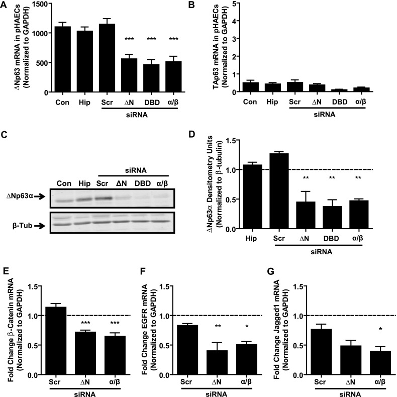
Figure 2.
Loss of p63 significantly down-regulates genes involved in epithelial function. (A) Quantitative RT-PCR of ΔNp63 mRNA in pHAECs, 48 hours after transfection with siRNA to N-terminally truncated (ΔN), DNA binding (DBD), or α/β domains of p63, indicates significantly decreased expression of ΔNp63 with each of the p63-specific siRNAs, but no change with HiPerfect (Hip) transfection reagent or scrambled siRNA (Scr) construct. Con, control. (B) Quantitative RT-PCR of TAp63 mRNA in pHAECs indicates no change at 48 hours after transfection with any siRNA constructs. (C) Representative immunoblot of ΔNp63α protein, 72 hours after transfection with 50 nM siRNA to ΔN, DBD, or α/β domains of p63. (D) Densitometry analysis of ΔNp63α expression in n = 3 pHAEC experiments. Values were normalized to β-tubulin as a loading control, and are shown as fold change over untreated control values (denoted with dashed line). (E–G) Quantitative RT-PCR of candidate p63 target genes by epithelial gene expression array, 48 hours after the knockdown of ΔN or α/β isoforms of p63 in n = 5 pHAECs. (E) β-catenin. (F) Epidermal growth factor receptor (EGFR). (G) Jagged1. Gene expression (relative to glyceraldehyde 3–phosphate dehydrogenase [GAPDH]) is shown as fold change over untreated control condition (dashed line). One-way ANOVA with the Dunnett post hoc test was used to assess statistical significance between treatments and scrambled siRNA control. *P < 0.05. **P < 0.01. ***P < 0.001.

