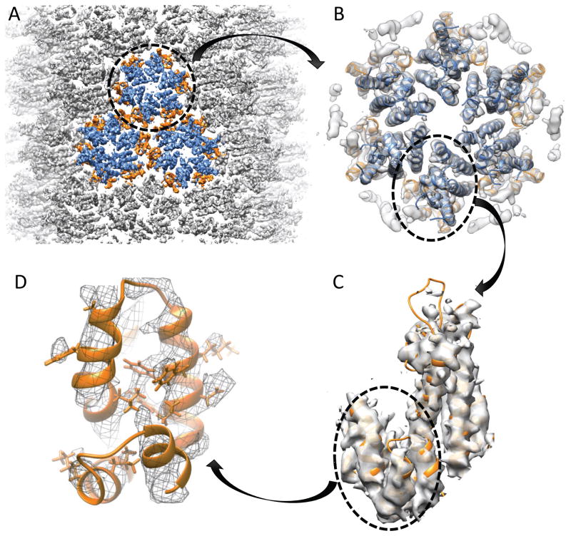
Figure 3.
CryoEM density map of the CA tubular assembly at 5 Å resolution, reconstructed from CA tubes with a helical symmetry of (−12, 11). A) Surface rendering of the density map of the CA tube, countered at 1.5σ. Three connecting CA hexamers are colored: CA-NTD; blue, CA-CTD; orange. B) A single CA hexamer is cut out from the lattice array, and MDFF fit using the crystal structure (PDB: 4XFX). C) Surface rendering of the CA monomer, cut out from the CA hexamer and countered at 1.2σ, displaying the helical turns for the alpha helices and some densities for the side chains. D) Surface mesh rendering of the CA-CTD, countered at 1.2σ, displaying the side chain densities of some bulky residues.

