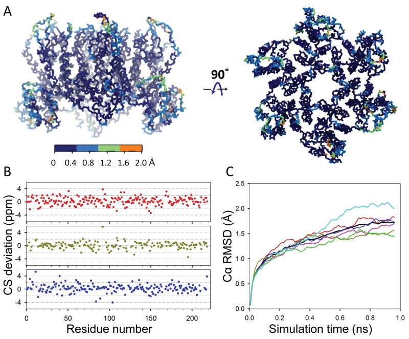
Figure 6.
MAS NMR CS-biased (backbone only) model of a CA hexamer in the CA tubular assembly. A) Root-mean-square fluctuations (RMSFs) resulting from the CS-biased simulation. Projection of the RMSFs on the structure of CA hexamer, ranging from 0 (dark blue) to orange (2.0 Å). B) Differences between the predicted CSs of the refined model and the experimental MAS NMR shifts for all CA residues, Cα in red, Cβ in dark green and C in blue. C) Time-evolution of the Cα RMSD between the CS-biased refined model and the starting MDFF model. All 6 CA chains are plotted in color, with the CA hexamer in black.

