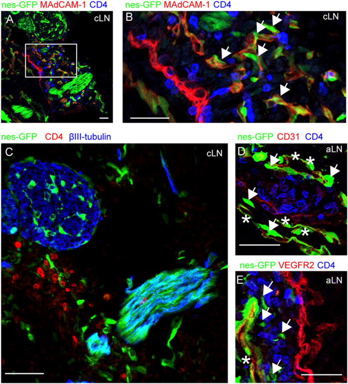
FIGURE 1.

Nestin-expressing stromal cells are present in E14.5 developing lymph nodes. (A–E) Immunofluorescence microscopy of lymph node anlagen of E14.5 nes-GFP mice. (A and B) Primitive lymph nodes were identified by CD4 (in blue), MAdCAM-1 (in red), and nestin-GFP cells (in green). (B) Magnification of a cervical lymph node anlage, containing MAdCAM-1+nestin-GFP+ cells (indicated with an arrow). (C) Cervical lymph node anlage found in close vicinity to bright GFP-expressing structures that coexpressed βIII-tubulin (in blue). (D and E) Axillary lymph nodes containing nestin-GFP cells that were either positive (asterisk) or negative (arrows) for CD31 (D, red) or VEGFR2 (E, red). Scale bars, 25 μm. Data are representative of four individual nestin-GFP embryos. aLN, axillary lymph node; cLN, cervical lymph node.
