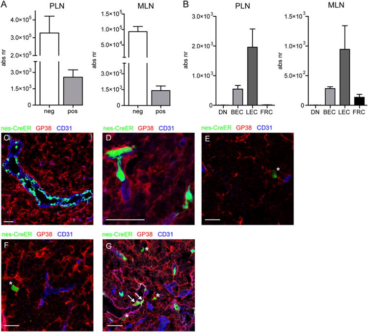
FIGURE 5.

During later stages of lymph node development, nestin predominantly labels endothelial cells. (A) Graphs showing the absolute number of GFP− and GFP+ cells of nes-creER c ROSA26-GFP mice that received tamoxifen at day 17 after birth and were analyzed 4 wk later. (B) Graphs showing the distribution of GFP+ cells among the various lymph node stromal populations. (C–G) Immunofluorescence analysis of nes-creER × ROSA26-GFP mice that received tamoxifen at p17 and were analyzed 4 wk later. (C–F) Lymph node sections stained for gp38 (red) and CD31 (blue) showing GFP+ HEVs (C), GFP+ capillaries (D), GFP+ gp38+CD31− stromal cells (indicated with asterisks, E–G), and GFP+gp38+ CD31+ lymphatic endothelial cells (indicated with arrows, G). Images are representative of lymph nodes of five individual animals (A and B) or three individual mice (C–G). Scale bars, 25 μm.
