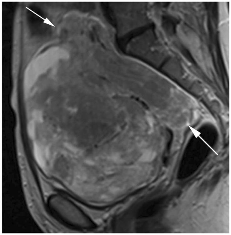
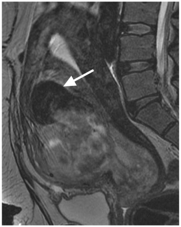
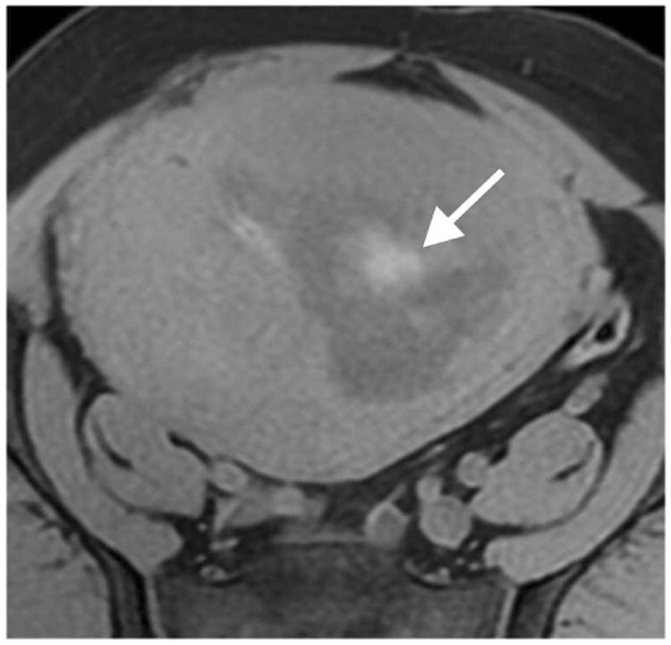
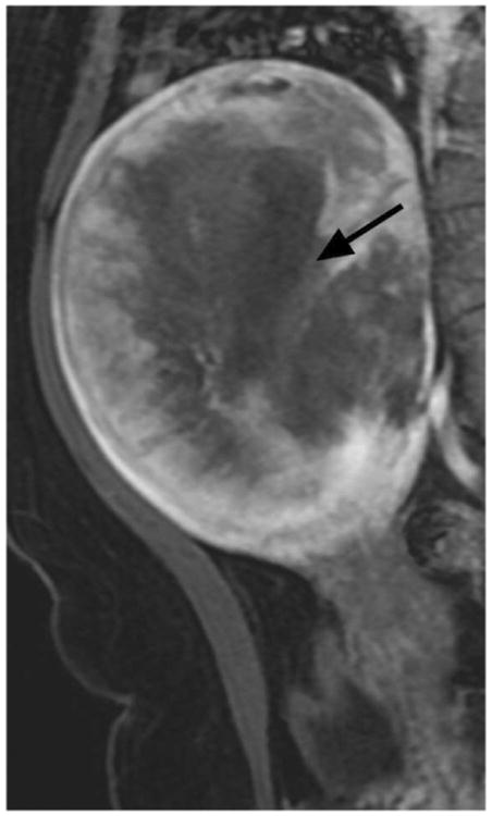
Figure 1.


Illustrations of the four qualitative MR features that demonstrated the strongest statistical associations with LMS at histopathology. A. Sagittal T2-weighted image shows a large uterine mass with nodular superior and posterior borders (white arrows). B. Sagittal T2-weighted image demonstrates “T2 dark” area in the myometrial mass (white arrow). C. Noncontrast T1-weighted fat-saturated image illustrates the presence of intra-lesional haemorrhage (white arrow). D. Sagittal contrast-enhanced T1-weighted fat saturated image shows the presence of central unenhanced areas (black arrow).
