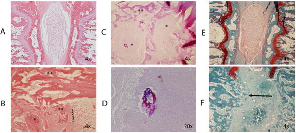
Figure 3.

A: H&E staining of control intervertebral segments. Normal architecture is preserved. B: H&E staining of infected intervertebral segment. Reactive bone formation (**), and abscess (*) are noted. Original position of endplate is noted (<). C: Gram staining of infected segment demonstrates abscesses (*) and reactive bone formation (**). D: Gram stain demonstrates bacteria in proximity to necrotic bone. E: Safranin-O and fast green staining of the control disc. F: Safranin-O staining of infected disc demonstrating endplate destruction and trabecular bridging (arrow). Remnants of the cartilaginous endplate are noted (++).
