
Fig. 1. Fig. 1.1: COS-7 cell transfected with dominant-negative (truncated) sortilin fused to a myc-tag protein.
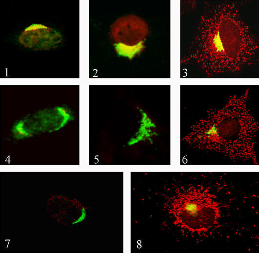
Anti-myc antibody (green) colocalizes with anti-Golgin antibody (red) in the perinuclear region of the cell. The overlay of both reactions yields yellow color indicating that truncated sortilin is retained in the Golgi apparatus. Magnification: x400. Fig. 1.2: COS-7 cell transfected with dominant-negative (truncated) sortilin fused to a myc-tag protein. Anti-myc antibody (green) colocalizes with anti-prosaposin antibody (red) in the perinuclear region of the cell. The overlay of both reactions yields yellow color indicating that prosaposin and sortilin were retained together in the Golgi apparatus. Magnification: x400. Fig. 1.3: COS-7 cell transfected with wild type sortilin construct (control) and immunostained with anti-Golgin (green) and anti-prosaposin (red) antibodies. The overlay shows yellow color in the perinuclear region and red granular in the cytoplasm typical of lysosomal staining. Magnification: x400. Fig. 1.4: COS-7 cell transfected with sortilin siRNA probe (sequence 1). The siRNA abolished the perinuclear staining of sortilin antibody (red). The overlay shows green fluorenscence only produced by the anti-Golgin antibody. Magnification: x400. Fig. 1.5: Cell transfected with a sortilin siRNA probe. The cell was stained with anti-sortilin (red) and anti-prosaposin (green) antibodies. The siRNA abolished the staining of sortilin and the punctate staining of the prosaposin antibody. Note that prosaposin is retained in the Golgi apparatus (green). Magnification: x400. Fig. 1.6: Control COS-7 cell (non transfected with siRNA) immunostained with anti-Golgin (green) and anti-prosaposin (red) antibodies. The red granular reaction is typical of prosaposin lysosomal staining. Magnification: x400. Fig. 1.7: COS-7 cell transfected with sortilin short hairpin interference RNA (shRNA) vector (sequence 1). The shRNA abolished the perinuclear staining of sortilin antibody (red). The overlay shows green fluorescence only, produced by the anti-Golgin antibody. Magnification: x400. Fig. 1.8: COS-7 cell transfected with empty vector (control) immunostained with anti-Golgin (green) and anti-prosaposin (red) antibodies. The red granular reaction is typical of prosaposin lysosomal staining. Magnification: x400.