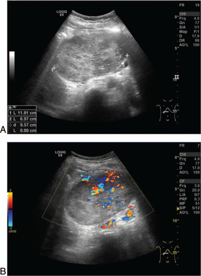
Figure 1.

Imaging studies using abdominal ultrasonography. (A) Ultrasonography showing an oval-shaped hypo- and isoechoic mass, 11.8 cm in size, located in the pancreatic head. (B) Visualization of the blood flow signal in and around the mass using color Doppler ultrasonography.
