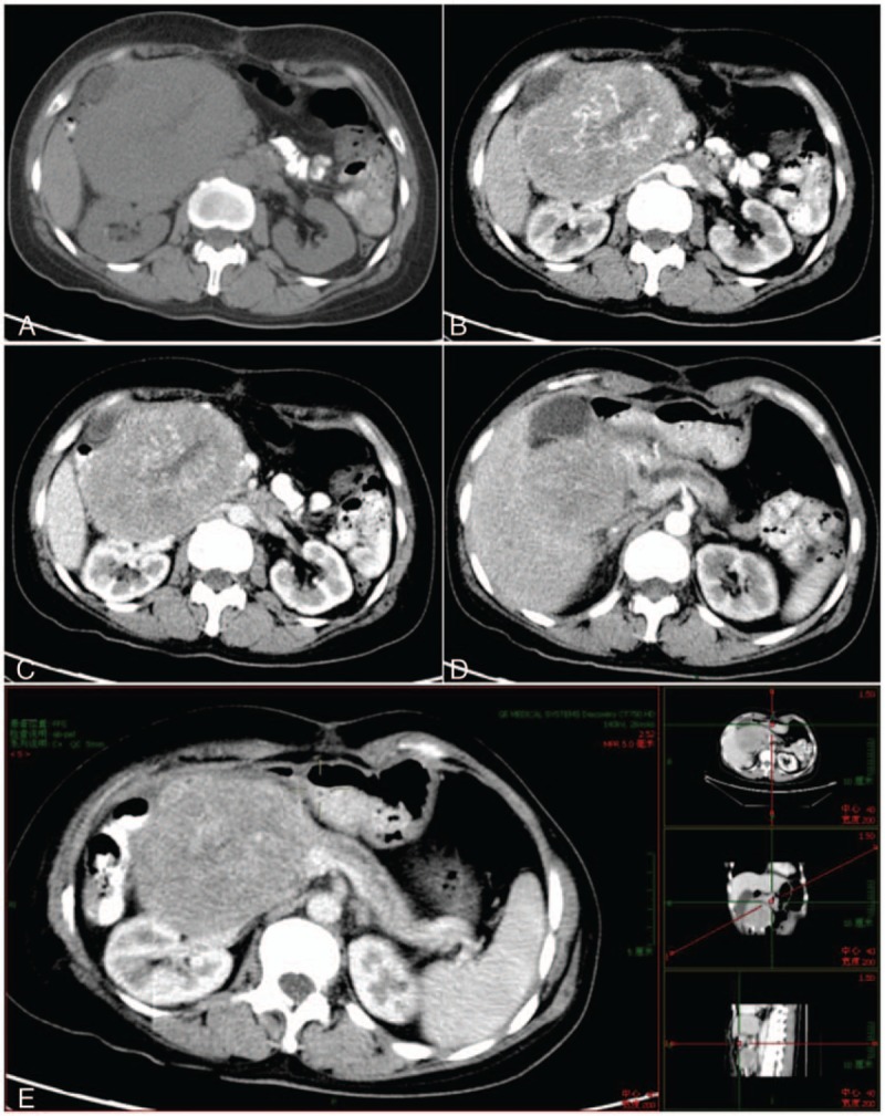
Figure 2.

Imaging of abdominal enhanced CT scans. CT showed a solid oval-shaped mixed-density mass, 11.5 cm in size, located in the pancreatic head. (A) Plain scan. (B) Arterial phase. (C) Vein phase. (D) The tumor invaded into the liver. (E) MPR revealed that the tumor was located in the pancreatic head. Dilatation of the upstream main pancreatic duct was also revealed. CT = computed tomography.
