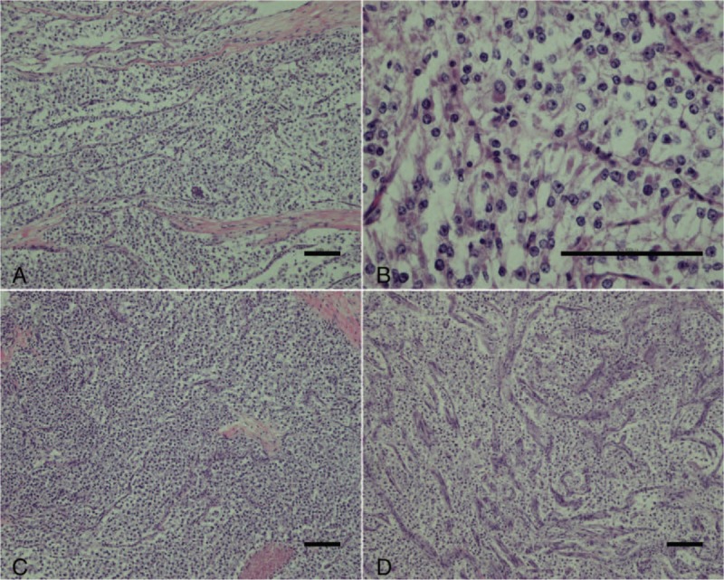
Figure 3.

(A,B) At the microscopic level, tumors were composed of epithelioid or spindle cells possessing clear to focally granular eosinophilic cytoplasm, centrally located round to oval nuclei, and inconspicuous nucleoli. (C, D) The tumor cells were arranged in nests or bundles. Some slender branching capillaries were revealed. (A–D: hematoxylin and eosin, scale: 100 μm).
