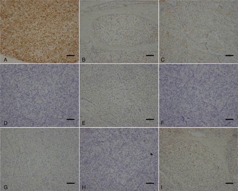
Figure 4.

Following immunohistochemical staining, related tumor cells were positive for human melanoma black 45 (A), progesterone receptors (B) and cluster of differentiation 56 (CD56) (C). However, these tumor cells were negative for Melan-A (D), S-100 (E), cytokeratin pan (F), chromogranin A (G), and CD10 (H). The Ki-67 labeling index was 5% (I) (scale 100: μm).
