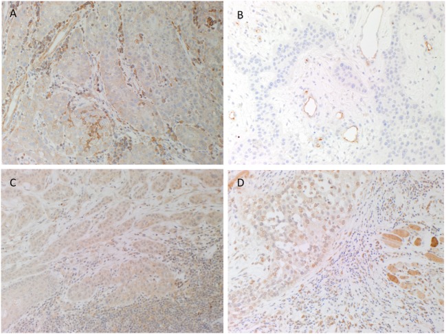
Figure 1.
Representative 3,3-diaminobenzidine immunohistochemical-stained sections of moderately differentiated lip cancer samples showing cytoplasmic expression of (pro)renin receptor [(A), brown] within the tumor nests (TNs) and cells within the stroma. Angiotensin-converting enzyme [(B), brown] was expressed on the endothelium of the microvessels within the stroma but not the TNs. Angiotensin II receptor 1 [(C), brown] showed weak to moderate cytoplasmic and nuclear staining by cells within the TNs and cells within the stroma. Strong perinuclear and cytoplasmic staining for angiotensin II receptor 2 [(D), brown] was present in cells within the TNs and the stroma, and skeletal tissue. Nuclei were counterstained with hematoxylin [(A–D), blue]. Original magnification: 200×.

