Figure 4:
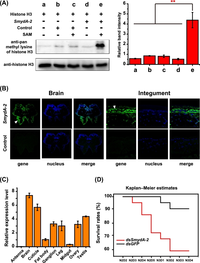
Function approval of SmydA-2 genes through experimental evidence. (A)In vitro methyltransferase assay of histone H3 of SmydA-2 in locusts. Anti-pan methyl lysine antibody recognizes histone H3 in vitro methylated with SmydA-2. Anti-histone H3 serves as endogenous control for protein samples. The analyses were carried out in three replicates. **P < 0.01. (B) Expression evidence of SmydA-2 in the brain and cuticle of locusts via fluorescence in situ hybridization analysis. Green signals indicate the expression of SmydA-2/control, and blue signals indicate nuclear staining with Hoechst. (C) Relative gene expression of SmydA-2 in the different tissues. mRNA levels are quantified using the SYBR Green expression assays on a LightCycler 480 instrument. The qPCR data are shown as the mean ± SEM (n = 6). (D) Survival analysis of the locusts after SmydA-2 double-strand RNA injection. Data are analyzed through the Kaplan–Meier survival curve comparison of the dsSmydA-2 and dsGFP groups for three replicates.

