
Figure 6.
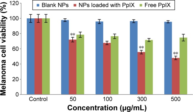
Cell viability of malignant melanoma A375 cells at different concentrations of blank nanoparticles, polymersomes loaded with PpIX, and free PpIX. Data are mean ± standard error of the mean; n=3. **P<0.01 compared to respective free PpIX. All values are different (P<0.01) from controls. For polymersomes loaded with PpIX, all values are different (P<0.01) at different concentrations.
Abbreviations: NPs, nanoparticles; PpIX, protoporphyrin IX.