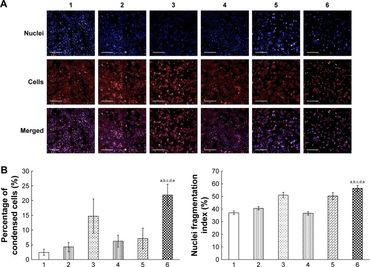
Figure 5.
Morphological changes of drug-resistant human breast cancer cells and their nuclei after treatment with various formulations.
Notes: Fluorescence images of the cells and their nuclei (A) were captured by a high-content screening system. The percentages of condensed cells (B) and the fragmentation index of nuclei (C) were calculated using the Columbus system. Drug-resistant human breast cancer MCF-7/adr cells were treated with blank medium (1), free epirubicin (2), free epirubicin plus quinine (3), epirubicin liposomes (4), combinational epirubicin liposomes (5), and functional epirubicin liposomes (6) for 12 h. The cells were then stained with Hoechst 33342 and Mitotracker Deep Red. In the combination formulations, the molar ratio of epirubicin to quinine was 1:1. Scale bar =200 μm. aP<0.05 versus blank medium; bP<0.05 versus free epirubicin; cP<0.05 versus free epirubicin plus quinine; dP<0.05 versus epirubicin liposomes; and eP<0.05 versus combinational epirubicin liposomes.

