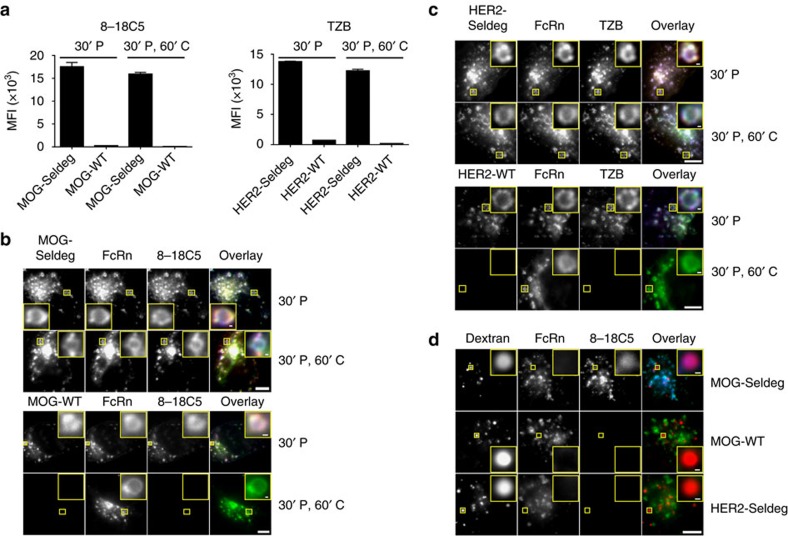
Figure 2. Seldegs increase the accumulation of antigen-specific antibodies in human endothelial (HMEC-1) cells expressing FcRn-GFP.
(a) HMEC-1 cells were incubated with 100 nM Alexa 647-labelled 8-18C5 (MOG-specific) or TZB (HER2-specific) in complex with 400 nM MOG-Seldeg/MOG-WT or HER2-Seldeg/HER2-WT for 30 min and chased for 0 (30' P) or 60 min (30' P, 60' C). Mean fluorescence intensities (MFI) of Alexa 647-labelled 8-18C5 or TZB for triplicate samples were determined by flow cytometry. Error bars indicate s.d. (b,c) HMEC-1 cells were plated on coverslips and treated as in a, except that Seldegs or control WT proteins were labelled with Alexa 555 and cells were fixed for microscopy. Images of representative cells from multiple cells analysed are shown with GFP, Alexa 555 and Alexa 647 in overlays pseudocoloured green, red and blue, respectively. Representative endosomes in the insets are cropped and expanded. (d) HMEC-1 cells were pre-pulsed with Alexa 555-labelled dextran for 2 h, washed and pulsed with 8-18C5 in complex with MOG-Seldeg, MOG-WT and HER2-Seldeg (concentrations and labels as for a) for 30 min, followed by an 8 h chase. Cells were washed, fixed and imaged, and images for a representative cell from multiple cells analysed are presented. Representative lysosomes in the insets are cropped and expanded. For the overlay, GFP, Alexa 555 and Alexa 647 are pseudocoloured as in b. For b–d, scale bars=5 μm, and for insets, scale bars=0.25 μm. Data shown are representative of at least two independent experiments.

