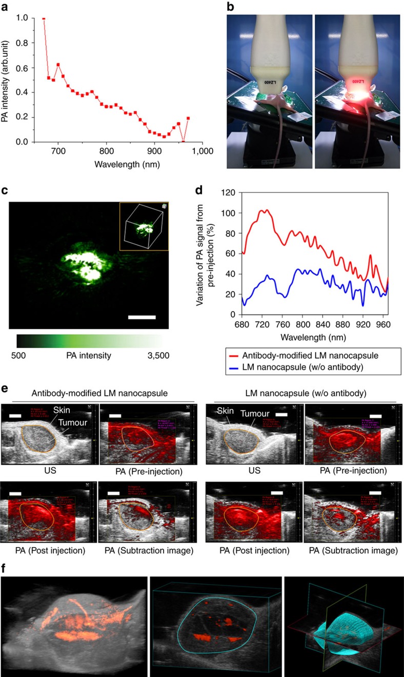
Figure 10. In vivo PA imaging using LM nanocapsules.
(a) PA intensity of LM nanocapsules (100 μg ml−1) at different wavelength. (b) Photos of in vivo PA imaging set-up before (left) and after laser exposure (right). (c) PA (green) image of LM nanocapsules (100 μg ml−1) under a mouse's skin. Excitation wavelength of laser is 680 nm. Inlet shows the 3D image of subcutaneous localization of LM nanocapsules. Scale bar, 4 mm. (d) Enhancement of PA intensity in tumour by antibody-functionalized LM nanocapsules (100 μg ml−1) in wide wavelength range for laser excitation. Variations of PA signals were calculated as post-injections minus pre-injections. (e) Antibody-functionalized LM nanocapsules (100 μg ml−1) targeted tumour imaging in living mice. Ultrasound (US) (grey) and PA (red) images were taken through the tumour by 750 nm laser excitation. Orange circles show the analysing parts of the PA signal for Fig. 8d. Scale bars, 2 mm. (f) 3D imaging of tumour treated by antibody-functionalized LM nanocapsules. Excitation wavelength of laser is 750 nm. Blue circle shows the part for construction of 3D structure. 3D, three-dimensional

