
Figure 4.
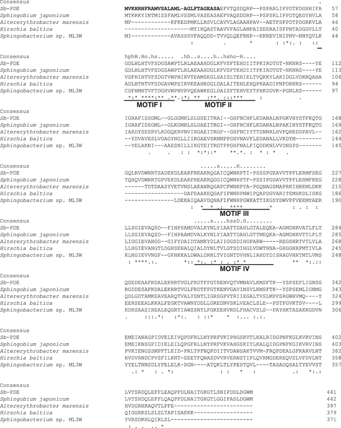
Sequence alignment of Sb-PDE with hypothetical proteins from Sphingobium japonicum (accession no. WP_006949123), Altererythrobacter marensis (WP_047806715), Hirschia baltica (WP_015828317) and Sphingobacterium sp. ML3W (WP_051959780). The numbers on the right are the residue numbers for each amino acid sequence. Identical residues and amino acid substitutions with low and high similarities are indicated by asterisks, dots and double dots, respectively. The putative signal peptide of Sb-PDE is in bold. The four conserved motifs in the PHP domain are underlined. The consensus sequence of each motif24 is shown above the alignment; h indicates hydrophobic residues (A, C, F, I, L, M, V, W or Y), s indicates small residues (A, C, S, T, D, N, V, G or P), p indicates polar residues (D, E, H, K, N, Q, R, S or T), o indicates hydroxy residues (S or T), and ‘−’ indicates negatively charged residues (D or E).