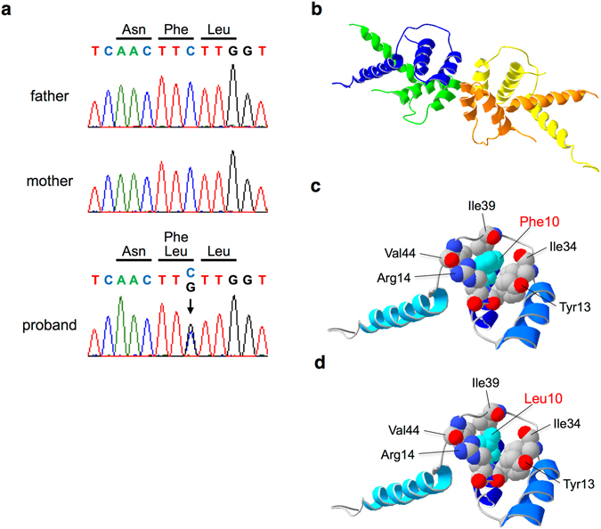
Figure 1.

Structural models of TBL1XR1 N-terminal domain (NTD). (a) De novo mutation in TBL1XR1 [c.30 C > G (p.Phe10Leu)]. The chromatogram shows the mutation in the TBL1XR1 gene, which is observed in the proband (arrow) but not in the parents. (b) Overall structure of a homology model of tetrameric NTD of TBL1XR1. Monomers are depicted in distinct colors. (c) A monomer model of the control NTD is depicted as a ribbon model. Important residues are depicted as spheres. Gray, red and blue spheres indicate carbon, oxygen and nitrogen atoms, respectively, although all atoms of Phe10 are colored in cyan for clarity. (d) A monomer model of Phe10Leu NTD is depicted as a ribbon model. Leu10 is colored in cyan for clarity.
