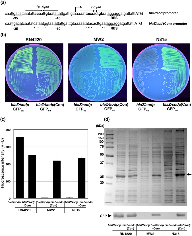
Figure 2.

Site-directed mutagenesis into the BlaI/MecI binding sequence. (a) Sequence alignment of the blaZ/sodp and its constitutively induced blaZ/sodp(Con) promoter region. Asterisks indicate the sequence exchanged by site-directed mutagenesis. Bold face indicates the BlaI/MecI binding motif (TACA/TGTA) within the larger palindromes, the R1 dyad and Z dyad are indicated by arrows. Up arrows with the tip to right indicates the transcription initiation site. Underlined sequences indicate −35 and −10 elements. Double underline indicates the RBS sequence. Initiation codons are shown in uppercase. (b) The fluorescing colonies were photographed under UV excitation. S. aureus RN4220, MW2 and N315 containing either pFK52, or pFK54 were grown on TSB agar plate containing chloramphenicol. (c) The comparison of fluorescence intensity among S. aureus strain RN4220, MW2, and N315 containing either pFK52, or pFK54. The fluorescent intensities at 513 nm were measured with a microplate reader, λex = 490 nm. The data represent mean values ± standard deviation. (d) The comparison of GFPsa expression efficiency among S. aureus strain RN4220, MW2, and N315 containing either pFK52, or pFK54. SDS-PAGE and Western blot analysis show the relative quantities of GFPsa in the whole cell lysates. The arrow indicates the position of GFPsa in gel. The Western blot gel was cropped and the full-length image is included in Supplemental Fig. 5(b).
