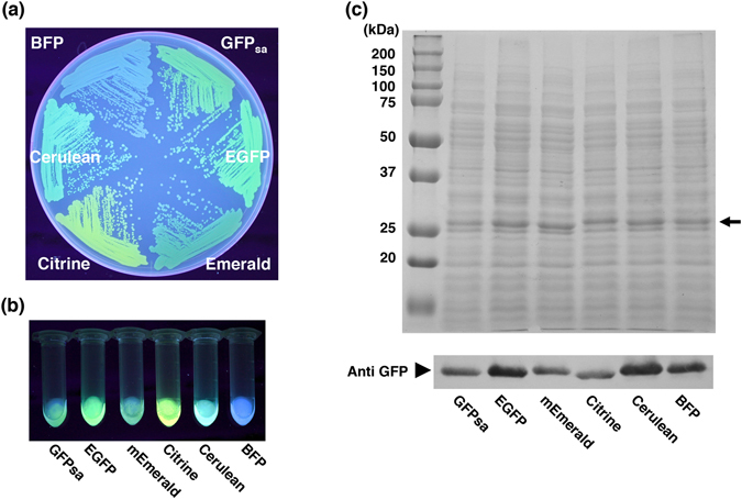
Figure 3.

Detection of multicolor GFP variants in the clinical strain, MW2. (a) The fluorescing colonies were photographed using UV excitation. S. aureus strain MW2 expressing multicolor GFP variants (GFPsa, EGFP, mEmerald, Citrine, Cerulean, and BFP) were grown on TSB agar plate containing chloramphenicol. (b) The whole cell lysates of S. aureus MW2 expressing multicolor variants used for SDS-PAGE and Western blot analysis were photographed under UV excitation. (c) The comparison of expression efficiency among multicolor GFP variants in S. aureus strain MW2. SDS-PAGE and Western blot analysis show the relative quantities of the multicolor GFP variants in the whole cell lysates. The arrow indicates the position of GFP variants in gel. The Western blot gel was cropped and the full-length image is included in Supplemental Fig. 5(c).
