Figure 2.

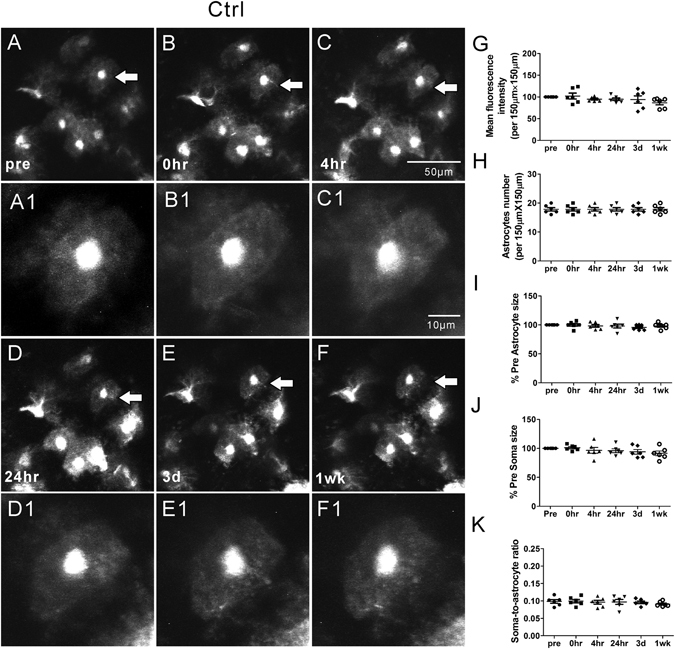
Representative in vivo images of astrocytes and quantitative analysis of astrocyte morphology features in the Ctrl group. Under normal physiological conditions, astrocytes typically have a characteristic bushy appearance consisting of thin process (A, A1). No obvious astrocytes vacuolization and morphological changes (B–F, B1–F1) were observed over a one-week period. No significant changes in mean fluorescence intensity (G), astrocyte number (H), astrocyte size (I), soma size (J) and soma-to-astrocyte ratio (K) occurred during a one-week period. (n = 6; one-way ANOVA or Kruskal-Wallis test, p > 0.05). The arrows in the lower magnification images indicate the astrocytes displayed in the higher magnification images.
