
Figure 4.
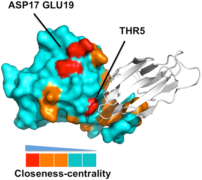
Surface representation of the garlic mannose-binding lectin protein (monomer, chain A). The residues are colored by closeness values with red, orange and cyan corresponding to the high (top 20%), intermediate (20~60%) and low (below than 60%) closeness values. The crystal structure of garlic lectin showed that THR5 is responsible for dimer formation and snowdrop lectin indicated that ASP17/GLU19 might be responsible for tetramer formation.