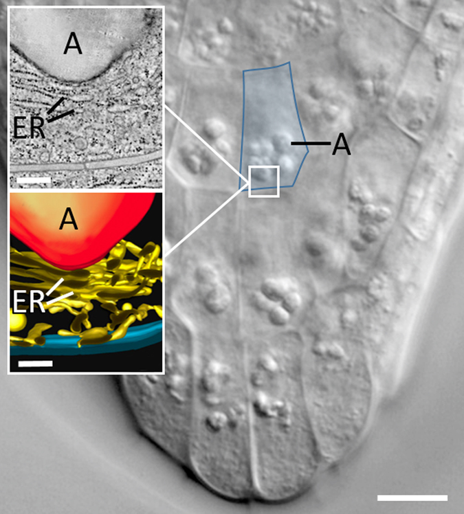
Fig. 3.

Differential interference contrast micrograph of a root cap of A. thaliana showing central columella cells with sedimented amyloplasts; a single columella cell is outlined in blue. Bar = 10 µm. Insets depict a tomographic slice image and a tomographic model of the lower part of an amyloplast (a) deforming tubules and cisternae of the endoplasmic reticulum network (ER) in the lower part of a columella cell of Medicago sativa. Bars = 300 nm. Modified after.60
