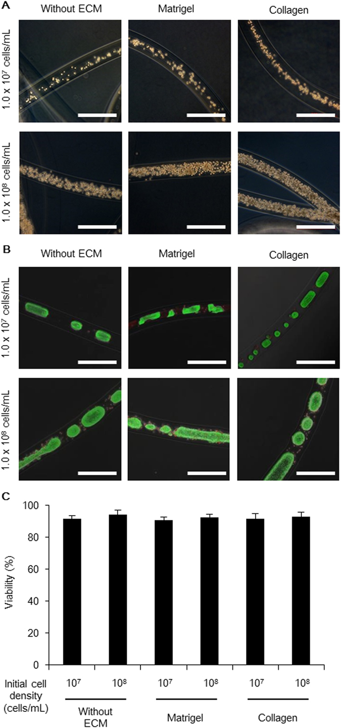
Figure 3.

Human iPS cells cultured in various types of core-shell microfibers. (A) Phase-contrast images showing the different types of human iPS cell-laden core-shell microfibers immediately after fabrication (day 0). Scale bar: 500 μm. (B) Merged phase-contrast and fluorescence images of the six types of core-shell microfibers cultured by day 4. Live cells were stained with calcein AM (green) and dead cells were stained with ethidium homodimer-1 (red). Scale bar: 500 μm. (C) Viability of the cells cultured in the six types of microfibers after the retrieval of cell aggregations from the microfibers and dissociation into single cells on day 4 (N ≥ 3).
