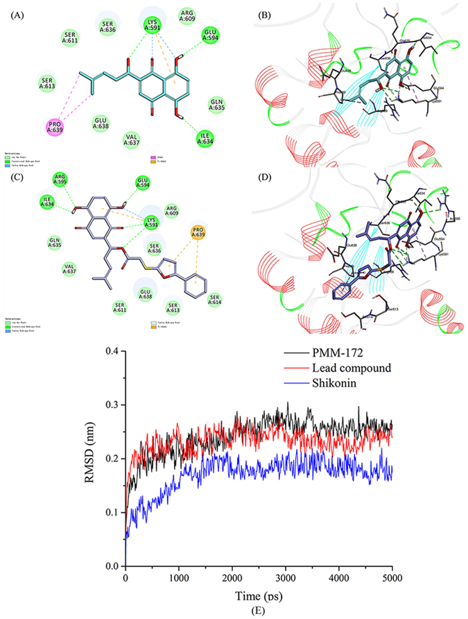
Figure 2.

Binding mode of SHK and lead compound PMM-158 with STAT3 (PDB code: 1BG1). (A) 2D diagram of the interaction between SHK and amino acid residues of the nearby active site. (B) 3D diagram of SHK inserted in the STAT3 binding site: for clarity, 3D structure of the protein is presented in line ribbon and only interacting residues are displayed. (C) 2D diagram of the interaction between PMM-158 and amino acid residues of the nearby active site. (D) 3D diagram of PMM-158 inserted in the STAT3 binding site: for clarity, 3D structure of the protein is presented in line ribbon and only interacting residues are displayed. (E)The RMSDs of the three studied complexes obtained during 5 ns of MD simulations, SHK (blue), lead compound (red) and PMM-172 (black).
