
Figure 4.
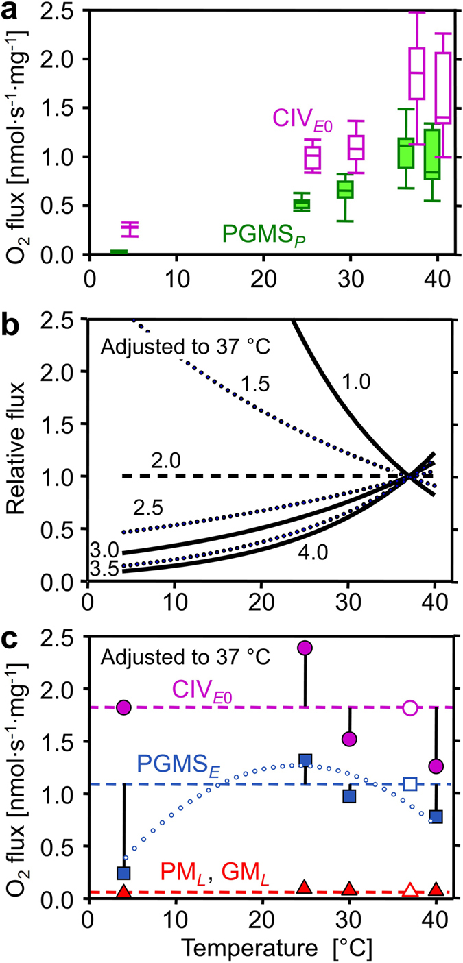
Effect of temperature on mitochondrial respiration. (a) Mass-specific oxygen flux (J O2) in respiratory states PGMSP (filled boxes) and CIVE0 extrapolated from the threshold plots in Fig. 5 (empty boxes). (b) Respiratory flux (j O2) relative to a simple temperature reference model: the reference flux is defined at 37 °C as 1.0 and at other temperatures as 1.0 if Q 10 is constant at 2.0 (horizontal dashed line). Non-linear deviations from standard conditions (full and dotted lines) are obtained when Q 10 differs from 2.0 (Q 10 shown by numbers) and is constant throughout the entire temperature range. (c) j O2 relative to standard temperature correction of flux at 37 °C (Q 10 = 2.0; horizontal lines): CIVE0 (● extrapolated from the threshold plots, Fig. 5), ETS capacity (■ PGMSE), and LEAK respiration (▲ pooled GML and PML). Vertical bars show deviations of experimental results (means, n = 5 to 14) from the theoretical line for a Q 10 of 2.0. The dotted trend line illustrates the change of temperature sensitivity for ETS, particularly at 4 °C. For box plots and abbreviations see Figs 1 and 2.