Abstract
Sarcomas are a heterogeneous group of rare tumors and arise either from soft tissue or from bone. Soft-tissue sarcomas (STSs) initially metastasize to the lungs. Metastases to extrapulmonary sites such as liver, brain, and soft tissue distant from primary tumor usually develop later. However, cases with isolated adrenal metastasis without disseminated disease have been reported in literature. We present a case of primary myxoid liposarcoma of the lower limb, in which staging 18-F fluorodeoxyglucose positron emission tomography-computed tomography (FDG PET-CT) scan detected a suspicious FDG avid adrenal lesion which eventually on resection was diagnosed as asymptomatic pheochromocytoma. Pheochromocytomas have been reported to demonstrate FDG uptake mimicking metastasis. Hence, while interpreting FDG PET-CT scans in the context of STSs, both the extrapulmonary metastatic potential of aggressive histological subtypes of sarcoma and rare possibility of FDG avid coexistent benign tumor should be taken into consideration.
Keywords: Adrenal lesion, 18-F fluorodeoxyglucose positron emission tomography-computed tomography, myxoid liposarcoma, pheochromocytoma
Introduction
Sarcomas, which account for <1% of cancers in adults, are relatively rare, and of those, about two-thirds arise from soft tissues and one-third from bone.[1] More than half of soft-tissue sarcomas (STSs) are found in the extremities, about 20% are found in the thorax, 15% are found in the abdomen, and 10% are found in the head and neck. Majority of extremity STSs initially metastasize to the lungs. Some metastases, called skip metastases, occur in soft tissue of the same limb as the primary tumor but at locations that are not adjacent to the primary tumor.[2] Extrapulmonary metastases usually appear after lung metastasis and represent disseminated disease. Initial metastases to other sites such as the liver, brain, and soft tissue distant from the primary tumor are rare.[3] However, isolated cases of adrenal metastasis without disseminated disease have been reported in the literature.[4,5] Pheochromocytomas are rare tumors of neuroectodermal origin arising from adrenal gland, and most tumors demonstrate uptake of 18-F fluorodeoxyglucose (FDG). Enhanced FDG uptake mimicking metastasis has been reported in benign pheochromocytomas without any correlation with catecholamine levels.[6] We present a case of primary myxoid liposarcoma of the lower limb with a suspicious FDG avid adrenal mass on FDG positron emission tomography-computed tomography (PET-CT) scan which on resection was diagnosed as asymptomatic pheochromocytoma.
Case Report
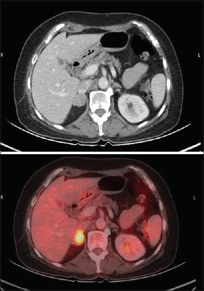
A 56-year-old female, known case of diabetes and hypertension for 2 years, well controlled with Telsartan-H and OHAs, presented with a large painless swelling in posterior aspect of the left thigh for 3 months, which was mobile from the underlying bone on clinical examination. Clinically, there was no evidence of neurovascular invasion and Tinel's sign was negative. Core biopsy of the lesion suggested tumor with myxoid background, foci of tumor necrosis, and few spindle cells with pleomorphic nuclei, immunonegative for CD34, EMA, S-100, SMA, and desmin suggesting myxoid liposarcoma (Ki67 75%). An FDG PET-CT scan from head to toe was carried out to accurately stage the disease. On the PET-CT scan, the large primary tumor involving posterior aspect of the left thigh measuring approximately 10.2 cm × 7.4 cm × 11 cm demonstrated moderate FDG uptake with a standardized uptake value (SUV) maximum of 5 [Figure 1]. There was no regional lymphadenopathy or lung nodule. However, an FDG avid lesion involving the right adrenal gland was seen demonstrating an SUV max of 9.4 [Figure 2]. Hence, the possibility of right adrenal metastasis though less likely was difficult to exclude. The patient was then worked up for possibility of pheochromocytoma and also planned for a laparoscopic right adrenalectomy for histological evaluation. Serum metanephrine, aldosterone, and cortisol levels were normal; however, plasma-free metanephrine levels were high-raising possibility of pheochromocytoma. However, in view of high FDG uptake and the patient being asymptomatic with well-controlled blood pressure, the possibility of metastases though rare was still difficult to rule out. Intraoperatively, the blood pressure of the patient was well maintained with uneventful intra- and post-operative period. The final histology of the adrenal mass showed a well-circumscribed nodular lesion with adjacent areas of hemorrhage. On microscopy, the tumor was composed of cells with finely granular amphophilic cytoplasm arranged in nested and trabecular pattern, positive for synaptophysin on immunohistochemistry and negative for vimentin and calretinin, suggestive of a pheochromocytoma. The patient is doing well on follow-up and is undergoing neoadjuvant radiation therapy to the primary STS prior to surgery.
Figure 1.

Whole body fluorodeoxyglucose positron emission tomography-computed tomography maximum intensity projection image demonstrating moderate-grade fluorodeoxyglucose uptake in primary soft-tissue sarcoma in the left thigh, and high-grade fluorodeoxyglucose uptake in right adrenal lesion (arrow)
Figure 2.

Contrast enhanced computed tomography transaxial slice and its corresponding fused positron emission tomography-computed tomography image demonstrating high-grade fluorodeoxyglucose uptake (standardized uptake value maximum 9.4) in right adrenal lesion measuring 2.4 cm × 2 cm
Discussion
STSs of the extremity are rare tumors, accounting for <1% of cancers in adults. Pulmonary metastases are common; however, extremity myxoid liposarcomas are known to have a unique extrapulmonary metastatic potential.[7] Isolated adrenal metastasis from primary STSs has previously been reported in literature without the development of pulmonary metastasis or disseminated disease. Kobayashi et al. have reported a case of bilateral adrenal metastasis with SUVs of 13.38 and 5.71 on FDG PET-CT without lung nodules at staging evaluation in a case of malignant fibrous histiocytoma of the buttock and thigh, and Mcphee et al. have reported a case of bilateral adrenal metastasis developing 16 months after treatment of primary cardiac sarcoma. Pheochromocytomas have been reported to demonstrate uptake of FDG with SUV maximum ranging from 1.9 to 42, with no statistically significant difference in SUV of metastatic versus nonmetastatic tumors.
Our case was an adult female who presented with large STS of the extremity, biopsy of which was suggestive of myxoid liposarcoma. FDG PET-CT scan demonstrated an FDG avid right adrenal mass which in view of the patient not having symptoms to suggest possibility of a pheochromocytoma and taking into consideration the extrapulmonary metastatic potential of myxoid liposarcomas, the probability of metastases was raised rather than a benign etiology. However, on further evaluation, the plasma-free metanephrines were found to be high and histologic evaluation of the adrenalectomy specimen was suggestive of pheochromocytoma.
Thus, our case highlights two important features that should be kept in mind while reporting FDG PET-CT scans in the context of STS: One, the extrapulmonary metastatic potential of certain histological subtypes of sarcomas and second, the possibility of FDG avid asymptomatic pheochromocytomas.
Conclusion
FDG PET-CT is an invaluable tool in oncology, especially for accurate staging of cancer to rule out metastatic disease. However, specificity remains a relative limitation as a host of benign lesions demonstrate FDG avidity. When the probability of metastatic lesion at a particular site is rare, the possibility of a benign condition even if not befitting the clinical scenario needs consideration.
Financial support and sponsorship
Nil.
Conflicts of interest
The authors declare no conflicts of interest.
Acknowledgment
We would like to acknowledge all the technical and supportive staff of Nuclear Medicine Department of P. D. Hinduja Hospital.
References
- 1.Altekruse SF, Kosary CL, Krapcho M, Neyman N, Aminou R, Waldron W, et al. SEER Cancer Statistics Review, 1975-2007. Bethesda, MD: National Cancer Institute; 2010. [Google Scholar]
- 2.Hosemann S. Stage II or III soft tissue sarcoma chemotherapy and radiation therapy before or after surgery benefit patients, OncoLog. Vol. 56. Houston, Texas: University of Texas M.D. Anderson Cancer Center; 2011. pp. 5–7. [Google Scholar]
- 3.Ryzewicz M, McLoughlin HA, Freudenberger C, Williams A, Lindeque B. Unusual metastases from extremity soft tissue sarcomas. Orthopedics. 2008;31:439. doi: 10.3928/01477447-20080501-25. [DOI] [PubMed] [Google Scholar]
- 4.Kobayashi E, Kawai A, Seki K, Tateishi U, Beppu Y. Bilateral adrenal gland metastasis from malignant fibrous histiocytoma: Value of [F-18]FDG PET-CT for diagnosis of occult metastases. Ann Nucl Med. 2006;20:695–8. doi: 10.1007/BF02984682. [DOI] [PubMed] [Google Scholar]
- 5.Rodríguez Corchero J, Medina López RA, Barrero Candau R, Agüelles Salido E, Pérez Espejo MP, Pascual del Pobil Moreno JL. Adrenal metastases of Ewing's sarcoma. Report of one case. Actas Urol Esp. 2004;28:70–4. doi: 10.1016/s0210-4806(04)73040-5. [DOI] [PubMed] [Google Scholar]
- 6.Taïeb D, Sebag F, Barlier A, Tessonnier L, Palazzo FF, Morange I, et al. 18F-FDG avidity of pheochromocytomas and paragangliomas: A new molecular imaging signature? J Nucl Med. 2009;50:711–7. doi: 10.2967/jnumed.108.060731. [DOI] [PubMed] [Google Scholar]
- 7.Estourgie SH, Nielsen GP, Ott MJ. Metastatic patterns of extremity myxoid liposarcoma and their outcome. J Surg Oncol. 2002;80:89–93. doi: 10.1002/jso.10093. [DOI] [PubMed] [Google Scholar]


