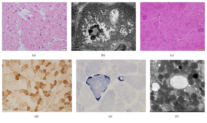
Figure 2.
(a) Light micrographs from endomyocardial biopsy revealed vacuolar change in myocardial cells, especially around nucleus and perimysial fibrosis without inflammatory cells (hematoxylin eosin stain, bar: 20 μm). (b) Electron microscopic examination of the endomyocardial biopsy revealed mitochondrial deformations with various types (e.g., sausage-like, loop-like, small, or gigantic), bar: 1 μm. (c) Light micrographs of the muscle biopsy revealed ragged red fibers (hematoxylin eosin stain, bar: 100 μm). (d) Light micrographs of the muscle biopsy revealed decreased cytochrome c oxidase (COX) activity (COX stain, bar: 100 μm). (e) Light micrographs of the muscle biopsy revealed blood vessels strongly reactive (SSV) to succinate dehydrogenase (SDH) (SDH stain, bar: 100 μm). (f) Electron microscopic examination of gastric biopsy revealed normal mitochondria (×1,2000).

