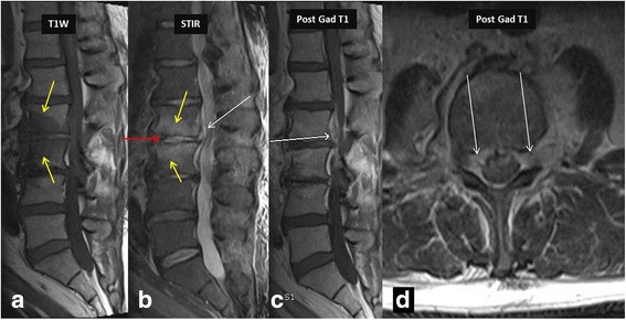
Fig. 2.

MR imaging in a 36-year-old intravenous drug abuser female shows marrow edema involving L2–3 vertebra reaching up to the endplates (yellow arrows in a and b), with minimal enhancement on post contrast imaging consistent with osteomyelitis. Note increased amount of fluid signal in L2–3 intervertebral disc suggesting discitis-osteomyelitis complex. Post contrast imaging shows enhancing epidural soft tissue (white arrows in b, c and d) without areas of liquefaction, consistent with epidural phlegmon. Once again post contrast imaging can differentiate epidural abscess from phlegmon and thus alter management. Also note the presence of left psoas muscle involvement. Responsible bacteria in this case were Staphylococcus epidermidis
