Fig. 1.
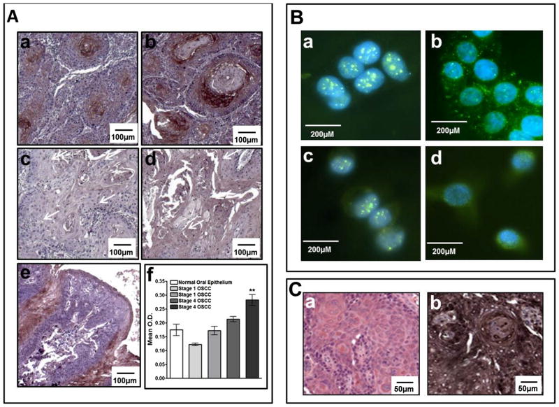
Panel A Immunohistochemical analysis of activated, phosphorylated ALK (phospho-ALK) in human OSCC tumors (4×). High phospho-ALK expression is illustrated in Stage IV OSCCs (panels a & b). Low phospho-ALK expression is illustrated in Stage I OSCCs (panels c & d). Normal tongue tissue (panel e) shows some artefactual staining due to tissue folding (panel e). Relative quantification is shown in panel f. Panel B: Immunofluorescent detection of total and activated (phospho-ALK) in OSCC cell lines (40×). Cal27 cells stained for total and phospho-ALK, panels a & b respectively; HSC3 cells stained for total and phospho-ALK, panels c & d respectively. Panel C: Immunohistochemical analysis of HSC3-derived tumors (20×). Representative H & E staining of HSC3-derived tumors is shown in panel a and corresponding phospho-ALK staining is shown in panel b.

