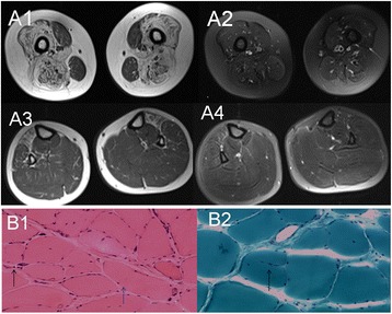
Fig. 4.

A1-4: The MRI of proband. A1-2 MRI revealed the bilateral thigh muscle disappear, the normal form was filled with a wide range of adipose tissue; A3-4 MRI showed the anteromedial calf group of muscles was infiltrated mild fatty. B1-2: Physical performance of proband; B1 (HE, ×100): the pathological changes in muscles showed that the sizes of muscle fibers were different, fiber atrophy (black arrow) and hypertrophy (blue arrow) alternately existed, compensatory hypertrophy of partial muscle fibers existed, fat and connective tissue proliferations were apparent. B2 (MGT, ×200): split muscle fibers can be seen in the hypertrophy muscle fibers (black arrow)
