
Fig. 1.
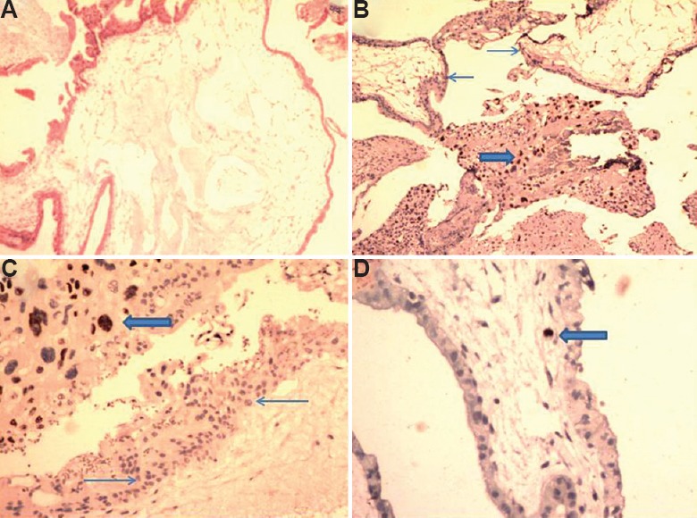
A case of complete mole showing (A) villous oedema with cistern formation (H & E, ×40); (B and C) p57 negativity in cytotrophoblast (thin arrows) with positivity in extravillous site (thick arrow; IHC, ×40 and ×100 respectively); (D) occasional stromal macrophage showing positivity (thick arrow; IHC, ×200).