Fig. 2.

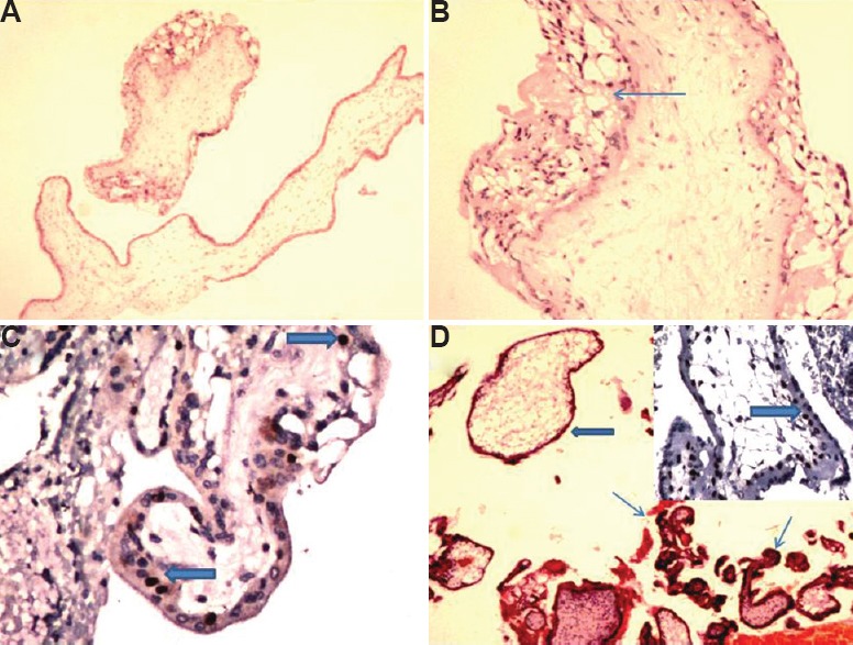
A case of partial mole showing (A) villous oedema with scalloping (H & E, ×40); (B) mild trophoblastic hyperplasia (thin arrow; H & E, ×100); (C) p57 positivity in cytotrophoblast(thick arrows; IHC, ×200); (D) case of hydropic abortus reclassified as partial mole showing a proportion of hydropic avascular villi (thick arrow) and a few normal appearing villi (thin arrows; H & E, ×40); inset showing p57 positivity in cytotrophoblast (thick arrow; IHC, ×200).
