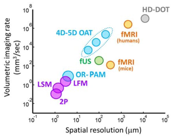
Fig 25.
Comparison of dynamic imaging capabilities of the various functional modalities used in small animal research and the clinics. Shown are: optical methods (violet) based on two photon microscopy (2P) [342], light-sheet microscopy (LSM) [343] and light field microscopy (LFM) [344]; small animal [345] and human [346] functional magnetic resonance imaging (fMRI - orange); high-density diffuse optical tomography (HD-DOT - gray) [347]; functional ultrasound (fUS - green) [348]; optical-resolution photoacoustic microscopy (OR-PAM) [22]; 4D and 5D optoacoustic tomography (4D-5D OAT) [255] (dots indicate three reported systems with isotropic resolution).

